

#Oct test registration
Once test registration is confirmed, there will be NO REFUNDS and you may not change your test, extend or cancel your registration.You must not use an account that does not belong to you for any reason. Each account can only register for one test at a time. You must be the person who registered the OCT account by which you use to register for and take tests.During an eye examination, you’ll have to stay still and look at the light from the equipment, whereas during a heart scan you will be under local anaesthetics which will avoid any pain.To ensure the integrity and fairness of online test, OCT has established and maintains the following rules and regulations: Test Rules 21-04-2016 30-03-2023Įdited by: Jay Staniland What is OCT (optical coherence tomography)? During an eye examination, you’ll have to stay still and look at the light from the equipment, whereas during a heart scan you will be under local anaesthetics which will avoid any pain. What does it feel like during the procedure? Therefore, you should not take anticoagulants, to avoid potential complications during or after the procedure. On the other hand, when doing a heart OCT, you will be under local anaesthetic (the catheter will need to reach the area to be analysed). However, you won’t be allowed to wear contact lenses during the exam. You won’t need to use eye drops as the machine won’t physically touch your eyes. When doing an eye OCT there is no need for any kind of preparations. In cardiology, OCT is used to determine whether there are thromboses, plaque, and calcium build-up or if a stent was placed incorrectly (especially when trying to prevent atherosclerosis). In ophthalmology, OCT is used to diagnose and assess the advancement and stage of retinal conditions such as maculopathies, glaucoma, hereditary retinal dystrophies, vitreoretinal diseases and macular oedemas.

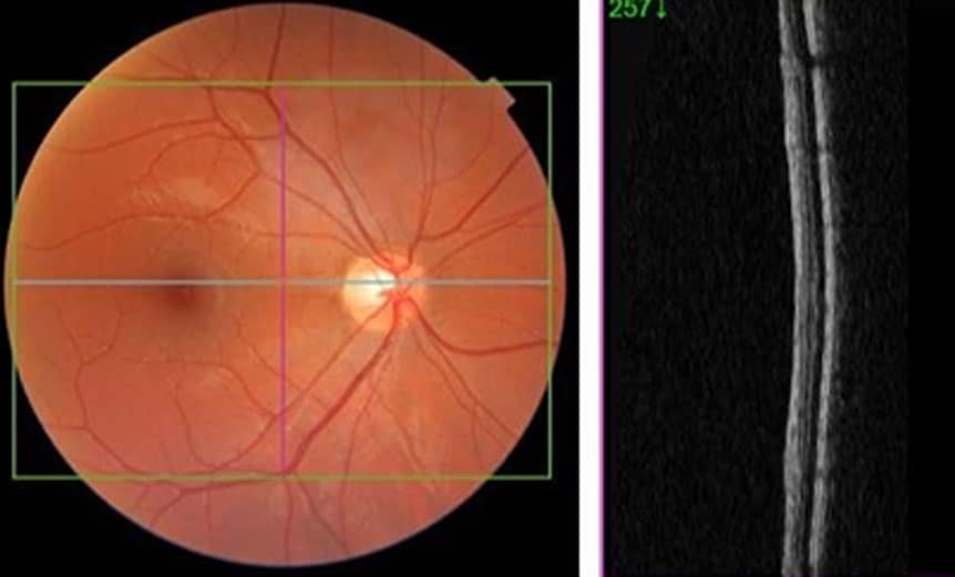
Once the light waves emitted by the transducer hit the clot and bounce back, they produce photon fluctuations which are sent back and received by an interferometer, allowing for an assessment of the problem. That means that the catheter has to physically reach the test area by making an incision. The result is an image of all the strata of the retina, allowing the specialist to determine if there are any abnormalities, from which they can formulate a diagnosis.įor cardiology purposes, this procedure is more invasive, as the light waves are sent from a catheter to the area of interest.

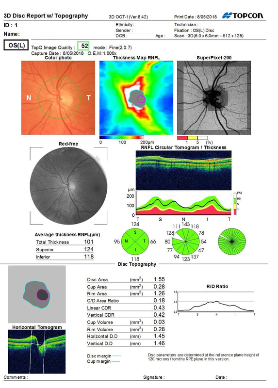
The test takes just a few minutes, during which the specialist will use certain reference points on the optic disk to receive and store the laser beams. Thanks to the possibility of analysing each tissue section, this exam can identify several conditions, especially those of the macula and of the optic disk. For ophthalmology purposes, this procedure is minimally-invasive, and it uses the infra-red laser rays to analyse the retina and cornea, generating precise and high definition pictures. This scan is primarily used in ophthalmology (to study the cornea and retina) and cardiology (to assess the state of the blood vessels where there may be a potential narrowing, which could lead to more severe complications).Īn OCT is similar to an ultrasound, however, light waves are used instead of ultrasound waves. OCT (optical coherence tomography) What is OCT (optical coherence tomography)?Īn optical coherence tomography (OCT) scan is an imaging test that uses low coherence light beams analyse different sections of the human body, obtaining highly precise and reliable pictures of several levels of tissue.


 0 kommentar(er)
